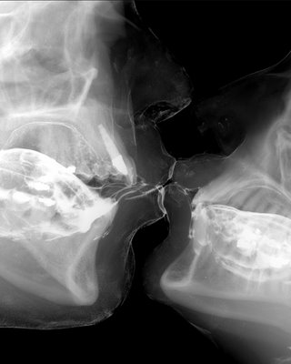
Enquanto existem pesquisadores tentando mostrar cientificamente o ato sexual, como neste trabalho, onde os autores utilizam o ultrassom 3d para demonstrar o coito: Deng, J., Hall-Craggs, M. A., Pellerin, D., Linney, A. D., Lees, W. R., Rodeck, C. H., et al. (2006). Real-Time three-dimensional ultrasound visualization of erection and artificial coitus. Int J Androl, 29(2), 374-9. Ou esses, que utilizam a ressonância magnética para o mesmo fim: Suh, D. D., Yang, C. C., Cao, Y., Heiman, J. R., Garland, P. A., & Maravilla, K. R. (2004). MRI of female genital and pelvic organs during sexual arousal. J Psychosom Obstet Gynaecol, 25(2), 153-62. Schultz, W. W., van Andel, P., Sabelis, I., & Mooyaart, E. (1999). Magnetic resonance imaging of male and female genitals during coitus and female sexual arousal. BMJ, 319(7225), 1596-600. Existem também artistas utilizando-se de métodos não convencionais para mostrar sua arte, como este que utiliza o Raio X tradicional para, também, mostrar o ato sexual: http://sex.innerpendejo.net/2007/10/wim-delvoye-sex-x-ray.html?zx=baaa8f…